
- The yeast-like colonies seen growing on the blood agar surface in the image below are lipid-dependent and grow only where olive oil had been applied. This is characteristic of the yeast-like organism, Malassezia furfur, which causes a superficial skin infection known as tinea versicolor.

Tinea Versicolor

Malassezia furfur Yeast in skin
- The lower image is a lactophenol blue mount made from a portion of the colony seen on the blood agar plate.
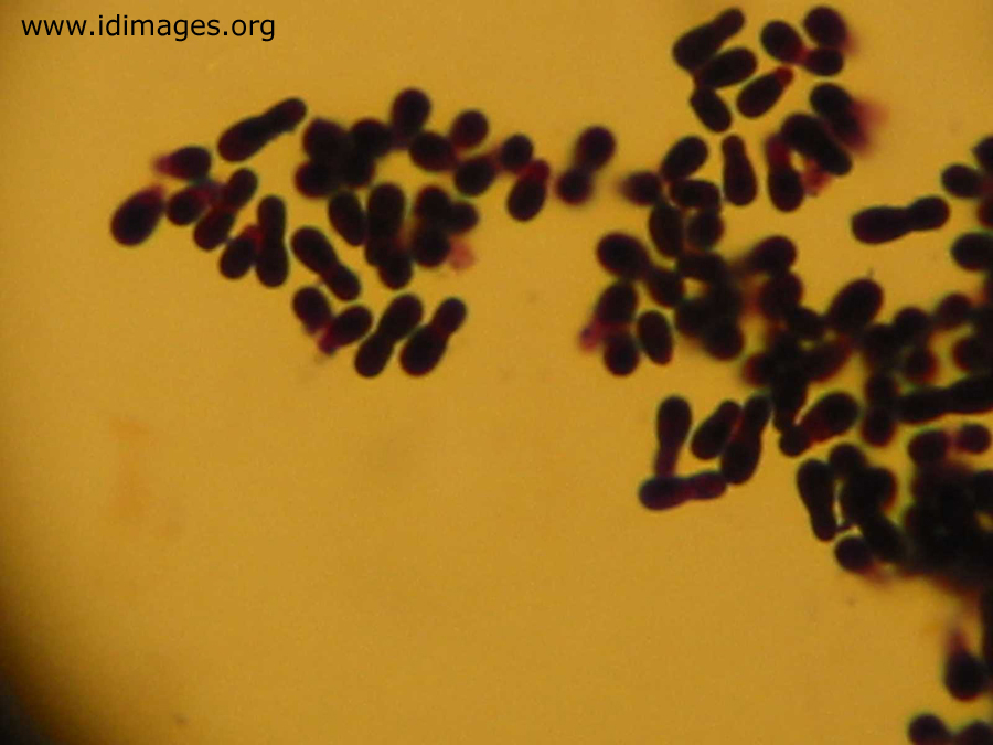
M. furfur stained with Lactophenol cotton blue

furfur – Broad based budding evident between daughter and parent yeast cell
Laboratory diagnosis:
- Clinical material: Skin scrapings from patients with superficial lesions, blood and indwelling catheter tips from patients with suspected fungaemia.
- Direct Microscopy: Skin scrapings taken from patients with Pityriasis versicolor stain rapidly when mounted in 10% KOH, glycerol and Parker ink solution and show characteristic clusters of thick-walled round, budding yeast-like cells and short angular hyphal forms up to 8um in diameter (ave. 4um diam.). These microscopic features are diagnostic for Malassezia furfur and culture preparations are usually not necessary.

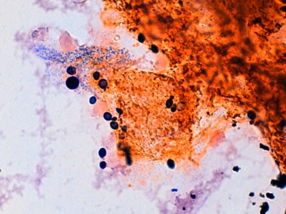
M. furfur KOH mount-preparation

M.furfur seen in a potassium hydroxide suspension. The appearance is that of typical yeast

M.furfur -arrows point to cells with broad-based budding
Microscopic Characteristics:
- Reproductive cell size is about 5 micrometers, with cells shaped like medicine capsules or sometimes bowling pins. Each produces a single phialoconidium followed by successive budding at a single location. Hyphae, very rarely produced in culture, are approximately 2 – 3 micrometers wide and short in length.
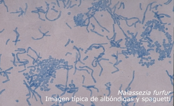
- In direct examination of a smear of infected tissue, hyphae are septate and hyaline usually not branched. Conidia resemble budding yeast cells and are approximately 3 micrometers in diameter. Together the hyphae and conidia are said to look like ‘spaghetti and meatballs.

Malassezia furfur under microscope from skin and agar culture
spaghetti and meatballs
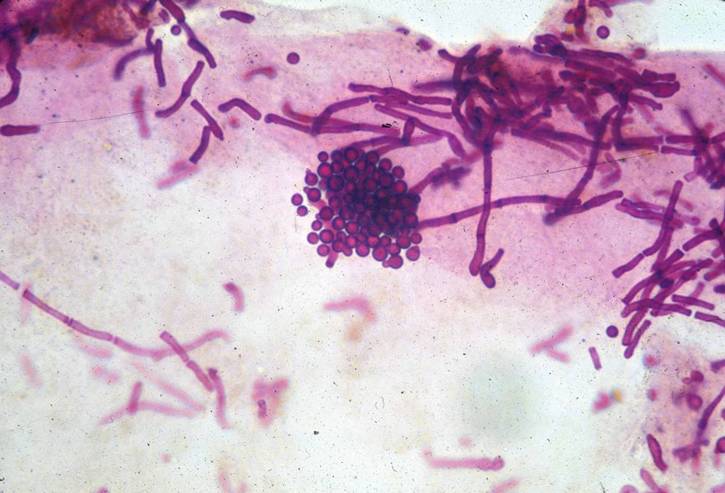
Gram stain of Malassezia furfur
M.furfur direct gram stain

Short blue-staining hyphae and spherical spores of Malassezia furfur
Macroscopic Characteristics:
- Slow growing colonies, appearing at temperatures of 35-37ºC. Colonies begin shiny and white to cream later becoming dull and beige, resembling bacteria-like colonies. Growth takes around one to two weeks on modified SDA which must be supplemented with fatty acids (usually done by covering medium with a thin layer of olive oil).

Malassezia furfur growing on a SDA with olive oil
- Malassezia species, M. furfur and M.pachydermatis, may also cause catheter related sepsis in patients receiving intravenous lipids.