
Principle
- Blood collected into an anticoagulant is placed in a long graduated tube held in a vertical position. The erythrocytes settle to the bottom, leaving a layer of plasma above.
- The height of the column of plasma after 1 hour indicates the sedimentation rate of the erythrocytes (erythrocyte sedimentation rate (ESR)).
Materials and reagents
- Westergren ESR tube: internal diameter 2.5mm; graduated from 0 to 200mm. (often marked 1 to 20: 1 corresponds to 10mm, 2 to 20mm, etc.)
- Westergren stand.
- Test-tubes.
- Graduated syringe, 5ml.
- Graduated pipette, 5ml.
- Anticoagulant: trisodium citrate, 3.2% solution (keep in refrigerator) or EDTA dipotassium salt, 10% solution.
Method
1. Pipette 0.4 ml of trisodium citrate solution into a test-tube or bottle.
2. Collect a venous blood specimen (see section 9.2). Apply the tourniquet as loosely as possible; puncture the vein at once and release the tourniquet. Collect 2 ml of blood into a syringe.
3. Remove the needle from the syringe and add 1.6 ml of blood to the test-tube containing anticoagulant (marked to contain a total of 2.0 ml) .Shakegently.
Measurement of the ESR should begin within 2 hours of collection of the blood.
4. Draw the citrated blood into the Westergren tube (using a rubber safety bulb) upto the 0-mm mark .
5. Place the tube in the Westergren stand, making sure that the tube is completely upright.
Check that there are no air bubbles in the tube.Check that the stand is level.
6. Leave on a bench away from vibration (e.g. not on the same bench as a centrifuge), free from draughts, not close to a central heating radiator and not in direct sunlight.
7. Wait 1 hour (set the timer to ring), then note the height of the column of plasma in mm graduations starting from the 0-mm mark at the top of the tube .
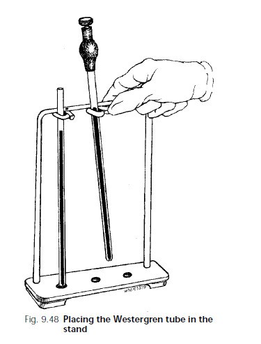
Placing Westergren tube in the stand
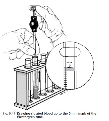
Westergren tube
Results
- The result is expressed in millimetres per hour (mm/h).
Reference range
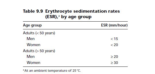
ESR Reference range by age
- Note: If a patient is dehydrated measurement of the ESR has little value.
High values
- Any disease that produces plasma protein changes will increase the ESR. These include acute and chronic infections, myocardial infarctions and rheumatoid arthritis.
An increased ESR rate may be due to:
- Anemia.
- Cancers such as lymphoma or multiple myeloma.
- Kidney disease.
- Pregnancy.
- Thyroid disease.
- autoimmune disorders include: Lupus ,Rheumatoid arthritis in adults or children.
Very high ESR levels occur with less common autoimmune disorders, including:
- Allergic vasculitis
- Giant cell arteritis
- Hyperfibrinogenemia (increased fibrinogen levels in the blood)
- Macroglobulinemia – primary
- Necrotizing vasculitis
- Polymyalgia rheumatica
An increased ESR rate may be due to some infections, including:
- Body-wide (systemic) infection
- Bone infections
- Infection of the heart or heart valves
- Rheumatic fever
- Severe skin infections, such as erysipelas
- Tuberculosis
Lower-than-normal levels occur with:
- Congestive heart failure
- Hyperviscosity
- Hypofibrinogenemia (decreased fibrinogen levels)
- Low plasma protein (due to liver or kidney disease)
- Polycythemia
- Sickle cell anemia
Reference:
– http://www.nlm.nih.gov
– Manual of basic techniques for a health laboratory, Second edition (World Health organization ,2003)