

Blood Plasma and Buffy Coat
Blood plasma:
is prepared by spinning a tube of fresh blood containing an anticoagulant (purple top tube) in a centrifuge until the blood cells fall to the bottom of the tube. The blood ...

Tips for making a good blood smear
<1> Clean slides
<2> The size of the drop: not more than 10microL
< 3> Speed of spreading action: Don’t move it too slow or too fast to have feathered edge smear.
<4> ...
Pappenheimer bodies VS Howell-Jolly bodies Differentiation
These inclusions are blue-purple in color, irregular in shape and located on the periphery of the cell. They were are Pappenheimer bodies. some people may think these inclusions ...

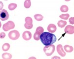
Howell and Jolly Body from Hair Cell Leukemia Patient
This arrow points to Howell and Jolly body, this blood smear has been taken from 86 old female patient presented with fatigue and abdominal pain, with increased white blood cell ...
sickle cell anemia and drepanocytes on smear
a 30 year-old female diagnosed with sickle cell anemia. “Sickle cell anemia (Hb SS disease) is caused by inheritance of two ßs genes and is the most serious of the sickling syndromes. ...

Infectious mononucleosis and atypical lymphocytosis on a smear
Infectious mononucleosis is an infection usually caused by the Epstein-Barr virus. The virus spreads through saliva, which is why it’s sometimes called “kissing disease.” ...

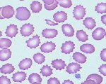
Crenated cells or Burr Cells
Crenated cell have pucker outer edges. Red cells are covered with 10 – 30 short spicules of regular form. (produced in a blood smear which dries slowly, also can be found in uremia, ...

Hypersegmented neutrophil with megaloblastic anemia on smear
Here is a hypersegmented neutrophil that is present with megaloblastic anemias. There are 8 lobes instead of the usual 3 or 4.
Such anemias can be due to folate or to B12 deficiency. ...

CBC diagram iron deficiency anemia
This is CBC diagram from patient with iron deficiency anemia, notice that:
Hemoglobin, Hematocrit is decreased —> Anemia
MCV is low —> Microcytic Anemia
MCH is low ...
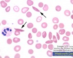
Aplastic Crisis in a Patient with Sickle Cell Disease
Numerous sickled RBC’s are present (small arrows).
A single nucleated RBC is noted (large arrow). Of note is the absence of polychromasia.

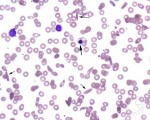
schistocytes and helmet cells in a smear accosiated with DIC
There are numerous fragmented RBC’s seen here. Some of the irregular shapes appear as “helmet” cells. Such fragmented RBC’s are known as “schistocytes” ...