


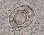
fertilized egg of Ascaris lumbricoides with Two divided cells
A fertilized egg of Ascaris lumbricoides.
Two divided cells are seen interior of the egg here.
Source: http://atlas.or.kr/
kidney bean-shaped of Balantidium coli cyst
Balantidium coli cyst in a stool specimen, a kidney bean-shaped macronucleus is observed.
The eggs of Ancylostoma and Necator
The eggs of Ancylostoma and Necator cannot be differentiated microscopically. The eggs are thin-shelled, colorless and measure 60-75 µm by 35-40 µm
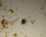
Hookworm egg in an unstained wet mount
Hookworm egg in an unstained wet mount, taken at 400x magnification.

Ascaris lumbricoides unfertilized eggs
Unfertilized eggs are elongated and larger than fertile eggs (up to 90 µm in length). Their shell is thinner and their mammillated layer is more variable, either with large protuberances ...

Eggs of Hymenolepis nana
These eggs are oval and smaller than those of H. diminuta, with a size range of 30 to 50 µm. On the inner membrane are two poles, from which 4-8 polar filaments spread out between ...
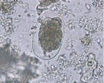
Artifact looks like the egg of Hymenolepis
Unknown object in a concentrated stool specimen. This object looks like the egg of Hymenolepis but lacks refractile hooks and the polar filaments seen in H. nana.


The eggs of Enterobius vermicularis
The eggs of Enterobius vermicularis measure 50-60 micron, are elongate-oval and slightly flattened on one side.
They are usually partially-embryonated when shed.
Enterobiasis can be ...

Trophozoite of E. histolytica n a direct wet preparation
Trophozoite of E. histolytica/E. dispar in a direct wet mount stained with iodine.

Trophozoites of E. histolytica with ingested erythrocytes stained with trichrome
Trophozoites of E. histolytica with ingested erythrocytes stained with trichrome.
The ingested erythrocytes appear as dark inclusions.
The parasites above show nuclei that have the ...