
Introduction:
- Entamoeba coli are a non-pathogenic ameba with world wide distribution. Its life cycle is similar to that of E. histolytica but it does not have an invasive stage and does not ingest red blood cells.
Morphology of Trophozoite:
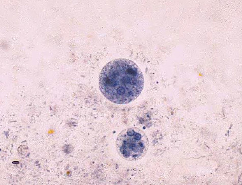
- The trophozoite is larger than that of E. histolytica ranging from 15-50μm in diameter. It exhibits blunt pseudopodia with sluggish movement. A permanently stained preparation shows a nucleus with a moderately large eccentric karyosome with the chromatin clumped on the nuclear membrane. The cytoplasm appears granular containing vacuoles with ingested bacteria and other food particles.
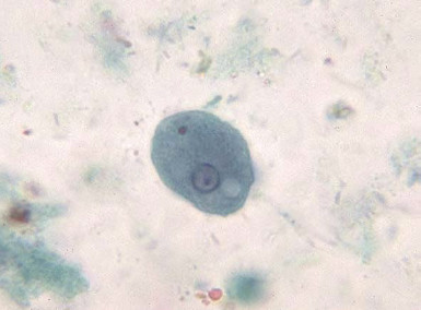
Entamoeba coli trophozoite with ingested bacteria

Entamoeba Coli Troph Pictures

Entamoeba coli Iodine stain trophozoite
Morphology of Cysts:
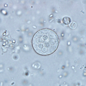
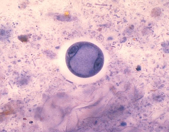
- Cysts of E. coli are 15-30μm in diameter and contain one to eight nuclei with irregular peripheral chromatin: karyosomes not central. Chromatoid bodies are not frequently seen but when present they are usually splinter-like with pointed ends. Glycogen is usually diffuse but in young cysts is occasionally found as a well-defined mass, which stains reddish brown with iodine.
Entamoeba coli (larger) and Entamoeba histolytica (smaller) cysts

Entamoeba coli (a) Trophozoite (b) Mature cyst with eight nuclei and splintered chromatoidal bars.
Cyst of Entamoeba coli in an unstained wet mount preparation with saline

Entamoeba coli. Two supernucleate cysts with 16 nuclei. Left, unstained cyst. Right, iodine-stained cyst.
Entamoeba coli: iron hematoxylin stain with Two mononucleate cysts: the large, central glycogen vacuoles have compressed the nuclei to the margin of the cyst
Laboratory Diagnosis:
- Laboratory diagnosis is made by finding the characteristic cysts in an iodine stained, formolether concentration method or by detecting the characteristic trophozoites in a wet preparation or a permanent stained preparation.
Microscopy:
- Where amebic dysentery is suspected, the laboratory should be informed that a “hot stool” is being supplied so that it can be examined within twenty minutes of being passed. On cooling the ameba stop moving which then become very difficult to identify. Direct microscopy should be done by mixing a small amount of the specimen in 0.9% sodium chloride solution. This permits detection of motile trophozoites of Entamoeba coli and can also provide information on the content of the stool (i.e., the presence of leucocytes and red blood cells). On search e.g.primarily for cysts, not for ameba, several stool samples are required to be examined, by direct microscopy and a sensitive concentration technique. Three negative stool samples are required before it can be accepted that there is no amebic infection. Microscopic examination of an amebic abscess aspirate (e.g. in the liver or lungs), may reveal hematophagous trophozoites. It must be examined immediately by mixing a drop of warm saline with some aspirated pus on a microscope slide.

Entamoeba coli. Wet mount examination of formalin-fixed specimen. Right, a trophozoite with hyaline ectoplasm (arrow) clearly distinct from the coarsely granular endoplasm containing numerous vacuoles; the nucleus is seen but it is usually not sufficiently distinctive for species identification. Left, only five nuclei are seen in this mature cyst. In formalin-fixed specimens, amoebic cysts (but not trophozoites) can be identified from the distinctive appearance of the nuclei.

entamoeba coli cysts – nuclei-chromatoid bars