

Platelets – what are they ?
Platelets:
Most are 1/5 to 1/3 the size of a normal RBC .
They are typically round with a blue-bray cytoplasm.
Cytoplasm with purple/blue granules.
Increased in:
Iron deficiency ...

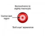
Codocytes known as Target Cells
Codocytes (“target cells”) are thin, hypochromatic cells with a round area of central pigmentation.
Codocytes are characteristically seen after splenectomy, and in patients ...

zone of morphology in a peripheral smear
A well-made peripheral smear is thick at the frosted end and becomes progressively thinner toward the opposite end. The “zone of morphology” (area of optimal thickness ...


relationship between HBV’s disease stages and AB-AG
A diagram shows the relation between HBV’s disease stages and AB-AG


Triple Phosphate Crystals
Triple Phosphate crystals are normal in urine
Appearance: typically appear in “coffin-lid” form. May also appear as “fern-leaf” shape if freshly formed.
Although ...

Calcium carbonate crystals
Calcium carbonate crystals are normal in urine
Appearance: small, colorless granules or dumbbells.
Not clinically significant but can be confused with other elements.
A unique feature ...
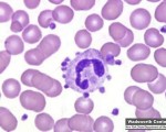
Platelet Satellitosis
Small platelets or platelet aggregates surrounding the cell membrane of a neutrophil or monocyte , this is called “Platelet Satellitism”
Found in:
EDTA in vitro induced ...

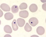
Howell-Jolly bodies in a thin blood smear stained with Giemsa
Howell-Jolly bodies are inclusion that may be seen in splenectomized patients or patients with an otherwise non-functioning or atrophic spleen, and in patients with severe anemia or ...