

Trophozoites of E. histolytica with ingested erythrocytes stained with trichrome
Trophozoites of E. histolytica with ingested erythrocytes stained with trichrome.
The ingested erythrocytes appear as dark inclusions.
The parasites above show nuclei that have the ...

Schistocyte in a case of β- thalassemia intermedia
This smear from a β- thalassemia intermedia patient, the arrow point to a Schistocyte
The Findings:
– Anisocytosis
– Poikilocytosis
– Hypochromia
– Target ...
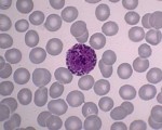
Basophil
Basophils are a specific type of white blood cell. These cells are readily stained with basic dyes (this is where the name comes from). Note the dark grains inside the cellular fluid ...

Band neutrophil or Staff Cell
Band neutrophil and also known as Staff Cell
2-3x larger than a mature RBC
Low nuclear to cytoplasmic (N/C) ratio (cytoplasm is relatively increased)
Kidney bean-shaped Nucleus with ...

Platelets – what are they ?
Platelets:
Most are 1/5 to 1/3 the size of a normal RBC .
They are typically round with a blue-bray cytoplasm.
Cytoplasm with purple/blue granules.
Increased in:
Iron deficiency ...


Calcium oxalate crystals
Calcium oxalate crystals (shown above) can be present in urine when oxalate-rich foods such as tomatoes, spinach, garlic, oranges, and asparagus are ingested.
Calcium oxalate ...
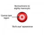
Codocytes known as Target Cells
Codocytes (“target cells”) are thin, hypochromatic cells with a round area of central pigmentation.
Codocytes are characteristically seen after splenectomy, and in patients ...

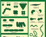
zone of morphology in a peripheral smear
A well-made peripheral smear is thick at the frosted end and becomes progressively thinner toward the opposite end. The “zone of morphology” (area of optimal thickness ...


relationship between HBV’s disease stages and AB-AG
A diagram shows the relation between HBV’s disease stages and AB-AG