
Featured Articles
 11 years ago
White blood cells: Description, Classification and Formation
 11 years ago
Blood Cells and Platelets
 11 years ago
Gram stain of wound specimen
All Stories

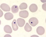
Howell-Jolly bodies in a thin blood smear stained with Giemsa
Howell-Jolly bodies are inclusion that may be seen in splenectomized patients or patients with an otherwise non-functioning or atrophic spleen, and in patients with severe anemia or ...
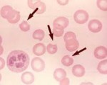
Bite cells known as Degmacytes
Bite cells (“Degmacytes”)
RBCs with peripheral single or multiple arcuate defects. Usually associated with spherocytes and blister cells.
seen in : Oxidant stress. Normal ...
coarse blue granules of Basophilic stippling
Basophilic stippling is the occurrence of fine, medium, or coarse blue granules uniformly distributed throughout some red blood cells.
Fine stippling may be associated with polychromatophilia, ...

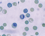
Reticulocytes using vital stain
In the presence of some anemias, the body increases production of red blood cells (RBCs), and sends these cells into the bloodstream before they are mature. These slightly immature ...

Giant Platelet on a smear
The arrow point to a Giant platelet, notice its size more than red cell size
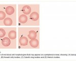
basophilic stippling – howel jolley bodies – Cabot Ring – Heinz bodies
A: Basophilic Stippling
B: Howel-Jolley Bodies
C: Cabot’s Ring
E: Heinz Bodies

Charcot-Leyden crystals
Charcot-Leyden crystals. These crystals are the breakdown products of eosinophils and maybe found in the feces and sputum of people with tissue-invading parasitic infections or various ...

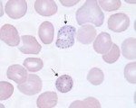
Cabot rings seen in megaloblastic anemia
Cabot rings are ring shape, or figure-8, these inclusions represents nuclear cabot rings can be seen in megaloblastic anemia

The rod-shaped appearance of the central pallor – stomatocyte
The rod-shaped appearance of the central pallor is characteristic of a stomatocyte.This image was obtained from the peripheral blood smear of an 86 year-old male who presented with ...